Our body is equipped with a protective skin barrier, the outermost layer of our body, often envisioned as a living wall of the body. This barrier protect us from environmental hazards, maintaining body integrity and survival. The formation/homeostasis of this protective layer involves contribution of the skin cells (keratinocytes, melanocytes, fibroblasts), and intracellular organelles/pathways. Using primary cells (healthy and diseased), organoid models, and employing cell biology and high-end microscopy approaches, we want to understand-
1.Skin Keratinocyte Differentiation:
Keratinocytes are the primary cell type in the epidermis, the outermost layer of the skin. They constitute 95% of the total cells and perform multiple functions. Keratinocytes mature through a process called differentiation that gradually transforms proliferative columnar cells (stem cells) into flattened, big sized differentiated cells. Differentiation ends with cornification of keratinocyte plasma membrane and cells are now enter into a state between life and death. These cells are now called corneocytes that are tightly packed together and constitute the skin barrier. They produce antimicrobial peptides, cytokines, and chemokines, which help to fight off infections and regulate inflammation. Corneocytes are sloughed off the skin on a monthly basis which renew the barrier. Keratinocytes act as sentinels, detecting pathogens and initiating immune responses. In most of the skin diseases keratinocyte differentiation is dysfunctional. Using cell biology/biochemistry and multi-omics approaches, we want to uncover the cellular processes/molecular mechanisms that act as a switch controlling this month-long gradual differentiation
Scientific References:
1.Mahanty S, Dakappa SS, Shariff R, Patel S, Swamy MM, Majumdar A, Setty SRG. Keratinocyte differentiation promotes ER stress-dependent lysosome biogenesis. Cell Death Dis. 2019;10(4):269.
2.Proksch E, Brandner JM, Jensen JM. The skin: an indispensable barrier. Exp Dermatol. 2008;17(12):1063-72.
3.Bikle DD, Xie Z, Tu CL. Calcium regulation of keratinocyte differentiation. Expert Rev Endocrinol Metab. 2012;7(4):461-72.

Differentiation of human primary keratinocytes: Cell size increases during early differentiation (at early stage), and form tissue with prolonged differentiation (Mahanty et al., 2019).
2.Endo-lysosomal pathway in Skin Epidermis Development:
During the gradual differentiation of keratinocytes, intracellular organelles—particularly those involved in the endo-lysosomal pathway (such as endosomes and lysosomes) and the secretory pathways (including the endoplasmic reticulum, Golgi apparatus, and plasma membrane)—undergo significant changes. We have demonstrated that the lysosomes in differentiated keratinocytes develop additional secretory properties, beyond their traditional role in degradation, which is typical of proliferative cells and other cell types. Using advanced microscopy techniques, including high-end and electron microscopy, along with various cell biology methods, we aim to understand the biological significance of organelle changes and, how associated molecular pathways adapt to facilitate these unique changes.
Scientific references:
1.Mahanty S, Dakappa SS, Shariff R, Patel S, Swamy MM, Majumdar A, Setty SRG. Keratinocyte differentiation promotes ER stress-dependent lysosome biogenesis. Cell Death Dis. 2019;10(4):269.
2.Mahanty S, Bergam P, Belapurkar V, Eluvathingal L, Gupta N, Goud B, et al. Biogenesis of specialized lysosomes in differentiated keratinocytes relies on close apposition with the Golgi apparatus. Cell Death Dis. 2024;15(7):496.
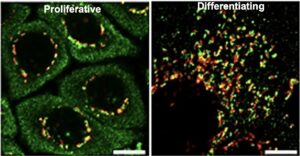
Re-distribution of the Golgi apparatus during keratinocyte differentiation.
Red = GM130; Green = p230. Scale bar = 10 μm.
A new class of specialized lysosomes are produced during keratinocyte differentiation. 3D rendering of the specialized lysosomes showing that lysosomes (in red) are enclosed by the Golgi protein GRASP65 (in green). Scale bar = 500 nm.
3.Skin Lamellar Body Biogenesis:
Skin lamellar bodies (LBs) are specialized organelles produced exclusively in keratinocytes. They are classified as lysosome-related organelles (LROs) and play a crucial role in forming the skin barrier. Unlike typical housekeeping organelles, such as lysosomes, mitochondria, the endoplasmic reticulum (ER), or the Golgi apparatus, LROs are specific to certain cell types. This means they are produced to fulfill particular functions in different tissues. For example, melanosomes in melanocytes provide photoprotection, lung lamellar bodies prevent lung collapse at the air-liquid interface, and Weibel-Palade bodies assist in blood clotting after injury. Skin LBs secrete lipids and a variety of antimicrobial peptides that envelop the corneocytes, forming a protective skin barrier. They also secrete proteases that facilitate desquamation, which is the renewal of the skin barrier. Dysfunction of lamellar bodies is associated with severe skin conditions, such as Harlequin Ichthyosis and atopic dermatitis. Our study will focus on the mechanisms of LB biogenesis, secretion, and their role in regulating epidermal immunity.
Scientific references:
1. Mahanty S SS. Epidermal Lamellar Body Biogenesis: Insight Into the Roles of Golgi and Lysosomes. Frontiers in Cell and Developmental Biology 2021;9:701950.
2. Mahanty, S., 2025. Skin lamellar bodies: a unique set of lysosome-related organelles. Frontiers in Cell and Developmental Biology, 13, p.1597696.
3.Feingold KR. Lamellar bodies: the key to cutaneous barrier function. Journal of Investigative Dermatology. 2012;132(8):1951-3.
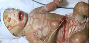
Harlequin Ichthyosis caused by lamellar body dysfunction.
Copyright: Wikipedia
B. Skin Barrier Regeneration and Disease Mechanisms:
We aim to uncover the role of skin immunity, sensation, and intercellular communication in skin regeneration after injury using laboratory-generated disease models and patient samples. Additionally, by modeling inflammatory diseases, we seek to investigate the role of keratinocytes in immune regulation and immune dysfunction.
Scientific references:
1.Tsepkolenko, A., Tsepkolenko, V., Dash, S., Mishra, A., Bader, A., Melerzanov, A. and Giri, S., 2019. The regenerative potential of skin and the immune system. Clinical, cosmetic and investigational dermatology, pp.519-532.
2.Abdo, J.M., Sopko, N.A. and Milner, S.M., 2020. The applied anatomy of human skin: A model for regeneration. Wound Medicine, 28, p.100179.

Different forms of psoriasis. Copyright: Wikipedia
C. Multi-scale model systems:
We are using below models to answer the above questions.
